Electron Microscopy Public Image Archive
Electron Microscopy Public Image Archive
大阪大学のEMPIAR-PDBjチームでは、アジアの電顕研究者が、EMPIARデータベースに大きな電顕画像を転送するお手伝いをしています。 ネットワークでイギリスに転送する代わりに、ハードディスク自体を郵送・宅配便で大阪大学に送付、あるいは阪大のサーバまでネットワーク転送していただければ、こちらで仲介して登録サイトへの転送を代行します。 EMPIARへの登録を希望する方で、阪大へのデータ送付・転送をご希望の方は、まず、e-mail で、登録したい電顕画像データについて、ご相談ください。
| Release date | Imageset | Title | Authors and references | Size | Resolution |
|---|---|---|---|---|---|
| 2023-01-19 | 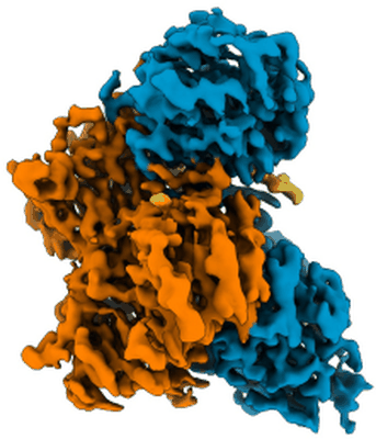 |
Human heparan sulfate polymerase complex EXT1-EXT2 [7046 micrographs in MRC format] | Leisico F, Omeiri J, Hons M, Schoehn G, Lortat-Jacob H, Wild R [Pubmed: 36402845] [DOI: 10.1038/s41467-022-34882-6] |
664.3 GB | 2.8 Å |
| 2023-01-18 | 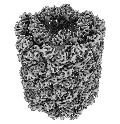 |
Cryo iDPC-STEM single particle analysis of keyhole limpet hemocyanin [multiple data sets in TIFF format] | Mann DM, Lazic I, Wirix M, de Haas F [Pubmed: 36064775] [DOI: 10.1038/s41592-022-01586-0] |
46.1 GB | 6.51 Å |
| 2023-01-18 | 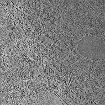 |
Tilt series of cell-cell contact of two PTK-1 cells [35 tilt series in MRC format] | Lemos M, Bezault A, Sauvanet C, Hanein D, Volkmann N [Pubmed: 36539423] [DOI: 10.1038/s41467-022-35409-9] |
1.2 GB | — |
| 2023-01-18 | 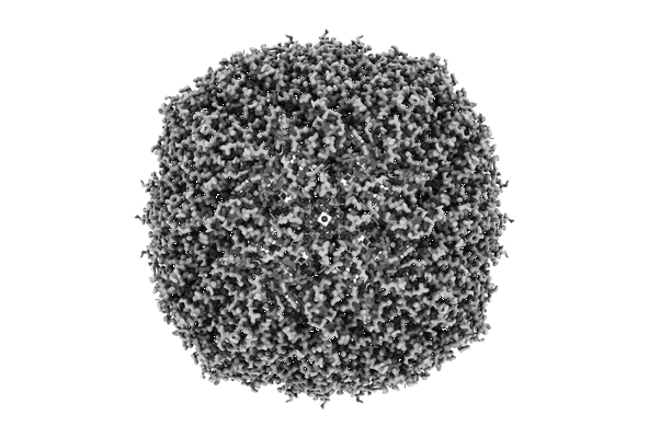 |
Movies of apoferritin collected at different dose rates on the Direct Electron Apollo direct detector - 30 eps [1478 multi-frame micrographs composed of 40 frames each in TIFF format] | Peng R, Fu X, Mendez JH, Randolph PS, Bammes BE, Stagg SM [Pubmed: 36578473] [DOI: 10.1016/j.yjsbx.2022.100080] |
374.2 GB | — |
| 2023-01-18 | 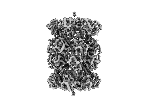 |
Multishot Tomography for High-Resolution In Situ Subtomogram Averaging: RiboProt singleshot [39 tilt series in MRC format] | Khavnekar S, Erdmann PSE, Plitzko J [Pubmed: 36343843] [DOI: 10.1016/j.jsb.2022.107911] |
131.2 GB | 4.7 - 7.8 Å |
| 2023-01-18 | 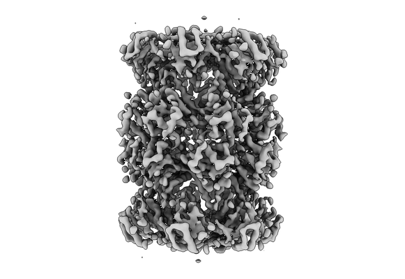 |
Multishot Tomography for High-Resolution In Situ Subtomogram Averaging: RiboProt multishot (2 shots) [26 tilt series in MRC format] | Khavnekar S, Erdmann PSE, Plitzko J [Pubmed: 36343843] [DOI: 10.1016/j.jsb.2022.107911] |
83.7 GB | 4.7 - 8.3 Å |
| 2023-01-18 | 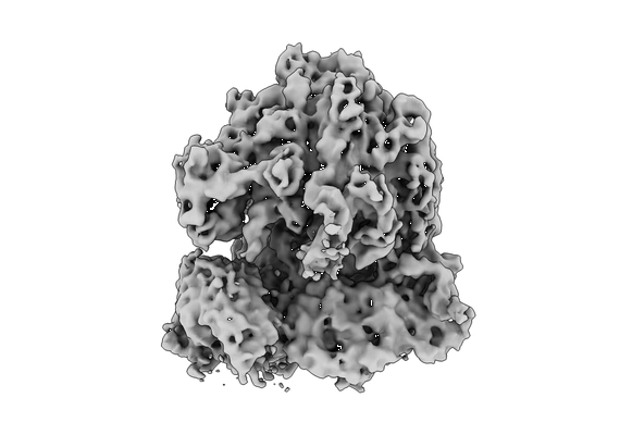 |
Multishot Tomography for High-Resolution In Situ Subtomogram Averaging: E.coli cryo-FIB lamellae multishot [30 tilt series in MRC format] | Khavnekar S, Erdmann PS, Plitzko JM [Pubmed: 36343843] [DOI: 10.1016/j.jsb.2022.107911] |
97.4 GB | 8.8 Å |
| 2023-01-18 | 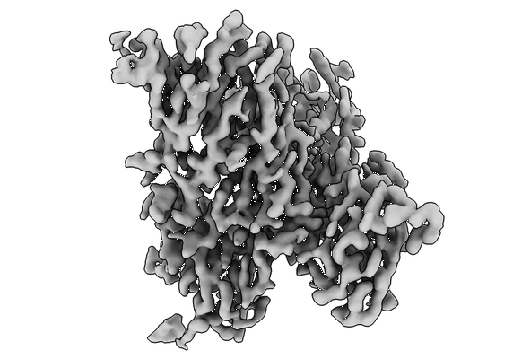 |
Organizing Structural Principles of the Interleukin-17 Ligand-Receptor Axis [multiple data sets in TIFF format] | Caveney NA, Wilson SC, Garcia KC [Pubmed: 35863378] [DOI: 10.1038/s41586-022-05116-y] |
19.5 TB | 2.5 - 4.4 Å |
| 2023-01-17 | 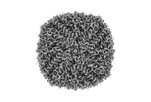 |
Movies of apoferritin collected at different dose rates on the Direct Electron Apollo direct detector - 60 eps [869 multi-frame micrographs composed of 20 frames each in TIFF format] | Peng R, Fu X, Mendez JH, Randolph PS, Bammes BE, Stagg SM [Pubmed: 36578473] [DOI: 10.1016/j.yjsbx.2022.100080] |
165.9 GB | 1.68 Å |
| 2023-01-16 | 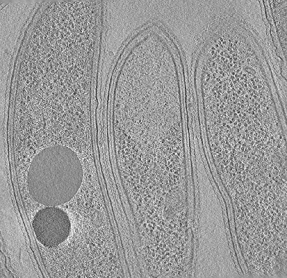 |
Cryo-electron tomography of FIB-milled Caulobacter crescentus expressing PopZ with IDR-156 and pentavalent OD [multiple data sets in MRC and TIFF formats] | Lasker K, Lam V, Villa E [Pubmed: 36163138] [DOI: 10.1038/s41467-022-33221-z] |
6.4 GB | — |
| 2023-01-16 | 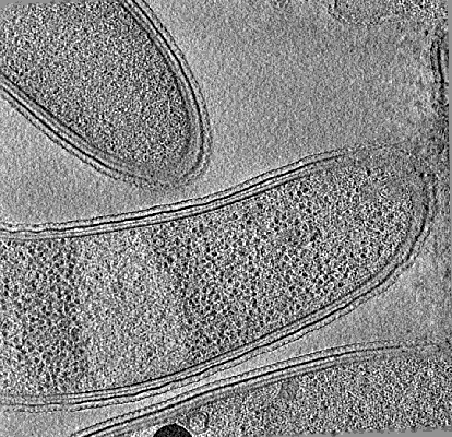 |
Cryo-electron tomography of FIB-milled Caulobacter crescentus expressing PopZ with IDR-156 [multiple data sets in MRC and TIFF formats] | Lasker K, Lam V, Villa E [Pubmed: 36163138] [DOI: 10.1038/s41467-022-33221-z] |
5.9 GB | — |
| 2023-01-16 | 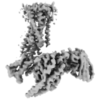 |
Structure of the active Gi-coupled human lysophosphatidic acid receptor 1 complexed with a potent agonist [6228 multi-frame micrographs composed of 48 frames each in TIFF format] | Akasaka H, Tanaka T, Sano FK, Matsuzaki Y, Shihoya W, Nureki O [Pubmed: 36109516] [DOI: 10.1038/s41467-022-33121-2] |
1.6 TB | 3.5 - 5.6 Å |
| 2023-01-16 | 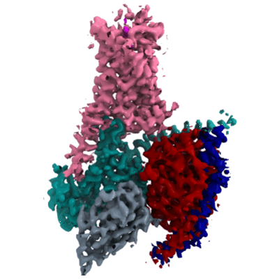 |
Cryo-EM structures of the β3 adrenergic receptor bound to solabegron and isoproterenol [multiple data sets in TIFF format] | Nagiri C, Kobayashi K, Tomita A, Kato M, Yamashita K, Nishizawa T, Inoue A, Shihoya W, Nureki O [Pubmed: 35489202] [DOI: 10.1016/j.bbrc.2022.04.065] |
2.8 TB | 3.3 - 3.9 Å |
| 2023-01-16 | 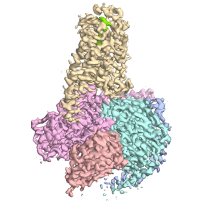 |
Dog beta3 adrenergic receptor bound to mirabegron in complex with a miniGs heterotrimer [2864 multi-frame micrographs composed of 48 frames each in TIFF format] | Nagiri C, Kobayashi K, Tomita A, Kato M, Yamashita K, Nishizawa T, Inoue A, Shihoya W, Nureki O [Pubmed: 34314699] [DOI: 10.1016/j.molcel.2021.06.024] |
720.6 GB | 3.16 Å |
| 2023-01-16 | 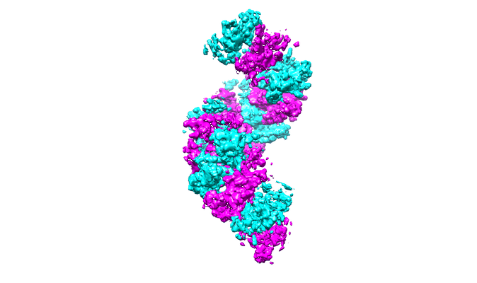 |
Structure of RecT protein from Listeria innoccua phage A118 in complex with 83-mer single stranded DNA [1619 multi-frame micrographs composed of 45 frames each in TIFF format] | Bell CE [Pubmed: 36543802] [DOI: 10.1038/s41467-022-35572-z] |
883.0 GB | 4.5 Å |
| 2023-01-10 |  |
Cryo-EM raw image of Bovine retinal pigmented epithelium lysate [multiple data sets in TIFF format] | Zhang Z., Morgan C.E. [Pubmed: 36577381] [DOI: 10.1016/j.celrep.2022.111876] |
2.0 TB | 2.28 - 3.32 Å |
| 2023-01-03 | 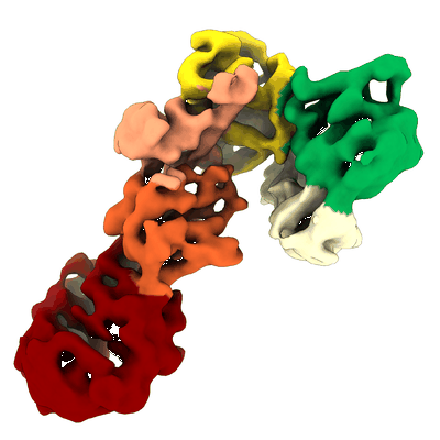 |
C-terminal half of Leucine Rich Repeat Kinase 1 (LRRK1) [3629 multi-frame micrographs composed of 50 frames each in TIFF format] | Matyszewski M, Leschziner AE [Pubmed: 36510024] [DOI: 10.1038/s41594-022-00863-y] |
1.3 TB | 5.8 Å |
| 2023-01-03 | 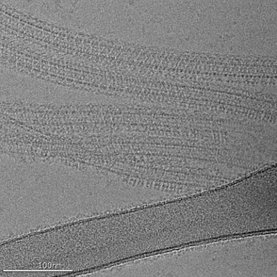 |
Apo C-terminal half of LRRK2 (I2020T) bound to microtubule [2379 multi-frame micrographs composed of 50 frames each in TIFF format] | Matyszewski M, Leschziner AE [Pubmed: 36510024] [DOI: 10.1038/s41594-022-00863-y] |
691.4 GB | 7.0 Å |
| 2023-01-03 | 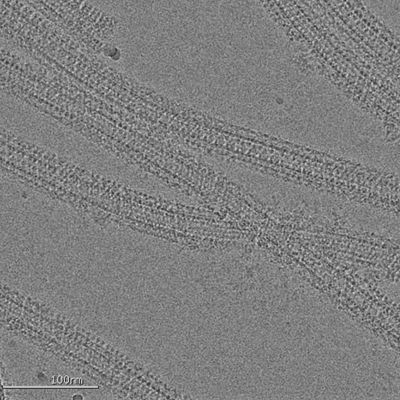 |
C-terminal half of LRRK2 (I2020T) bound to microtubule in presence of MLi-2 kinase inhibitor [2354 multi-frame micrographs composed of 55 frames each in TIFF format] | Matyszewski M, Leschziner AE [Pubmed: 36510024] [DOI: 10.1038/s41594-022-00863-y] |
735.3 GB | 4.5 - 18.0 Å |
| 2023-01-03 | 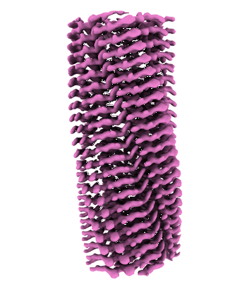 |
Cryo-EM structure of hnRNPDL amyloid fibrils [multiple data sets in TIFF format] | Chaves-Sanjuan A, Garcia-Pardo J, Bartolome-Nafria A, Gil-Garcia M, Visentin C, Bolognesi M, Ricagno S, Ventura S [Pubmed: 36646699] [DOI: 10.1038/s41467-023-35854-0] |
945.0 GB | 2.5 Å |
| 2023-01-03 |  |
Movies of apoferritin collected at different dose rates on the Direct Electron Apollo direct detector - 15 eps [972 multi-frame micrographs composed of 76 frames each in TIFF format] | Peng R, Fu X, Mendez JH, Randolph PS, Bammes BE, Stagg SM [Pubmed: 36578473] [DOI: 10.1016/j.yjsbx.2022.100080] |
319.1 GB | — |
| 2023-01-03 |  |
Cryo electron tomography of Ca. L. ossiferum [multiple data sets in MRC format] | Wollweber F, Xu J [Pubmed: 36544020] [DOI: 10.1038/s41586-022-05550-y] |
12.5 GB | 11.7 - 24.5 Å |
| 2022-12-19 |  |
CLEMSite, a software for automated phenotypic screens using light microscopy and FIB-SEM. [multiple data sets in TIFF format] | Lleti JMSL, Steyer AMS, Schwab YS | 21.2 GB | — |
| 2022-12-16 |  |
Amyloid fibril structure from the vascular variant of systemic AA amyloidosis [multiple data sets in TIFF format] | Banerjee S, Baur J, Daniel C, Pfeiffer PB, Hitzenberger M, Kuhn L, Wiese S, Bijzet J, Haupt C, Amann KU, Zacharias M, Hazenberg BPC, Westermark GT, Schmidt M, Fändrich M [Pubmed: 36433936] [DOI: 10.1038/s41467-022-34636-4] |
1.1 TB | 2.56 Å |
| 2022-12-13 | 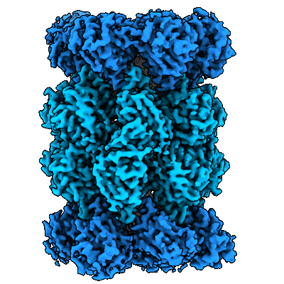 |
Single particle cryo-EM dataset of the Vairimorpha necatrix 20S proteasome from spores [7240 multi-frame micrographs composed of 30 frames each in TIFF format] | Jespersen N, Ehrenbolger K, Winiger RR, Svedberg D, Vossbrinck CR, Barandun J [Pubmed: 36379934] [DOI: 10.1038/s41467-022-34691-x] |
874.1 GB | 2.77 Å |
