Electron Microscopy Public Image Archive
Electron Microscopy Public Image Archive
The EMPIAR-PDBj team at Osaka University assists Asian EM researchers with the transfer of big EM image data to EMPIAR. Instead of sending the data directly to the EBI (UK) via the internet, hard drives can also be sent to Osaka University by postal mail or via a courier service. As an alternative, internet transfer to our server in Osaka is also available. If you would like to take advantage of our submission services, please contact us first by e-mail before sending the data to us.
| Release date | Imageset | Title | Authors and references | Size | Resolution |
|---|---|---|---|---|---|
| 2023-08-18 | 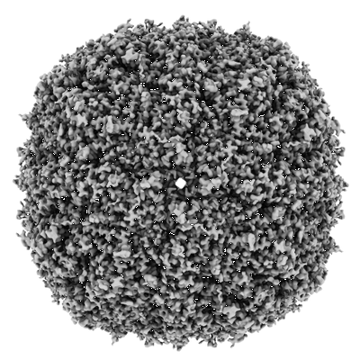 |
Mouse heavy chain apoferritin in vitreous ice after laser-melting and revitrification [12047 multi-frame micrographs composed of 553 frames each in EER format] | Bongiovanni G, Harder OF, Drabbels M, Lorenz UJ [Pubmed: 36438663] [DOI: 10.3389/fmolb.2022.1044509] |
2.2 TB | 1.63 Å |
| 2023-08-18 |  |
Initiation complex consisting of E. coli 70S ribosome with AAA mRNA codon in the A-site incubated with a ternary complex containing cognate acylated tRNA(Lys) [8435 multi-frame micrographs composed of 40 frames each in TIFF format] | Koziej L, Glatt S [Pubmed: 37553384] [DOI: 10.1038/s41467-023-40422-7] |
3.2 TB | 2.33 - 2.88 Å |
| 2023-08-18 | 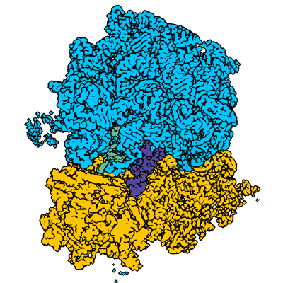 |
Initiation complex consisting of E. coli 70S ribosome with AAm6A mRNA codon in the A-site incubated with a ternary complex containing cognate acylated tRNA(Lys) [7207 multi-frame micrographs composed of 40 frames each in TIFF format] | Koziej L, Glatt S [Pubmed: 37553384] [DOI: 10.1038/s41467-023-40422-7] |
2.7 TB | 2.11 - 2.62 Å |
| 2023-08-18 | 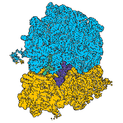 |
Initiation complex consisting of E. coli 70S ribosome with Am6AA mRNA codon in the A-site incubated with a ternary complex containing cognate acylated tRNA(Lys) [7172 multi-frame micrographs composed of 40 frames each in TIFF format] | Koziej L, Glatt S [Pubmed: 37553384] [DOI: 10.1038/s41467-023-40422-7] |
2.7 TB | 2.21 - 2.81 Å |
| 2023-08-18 | 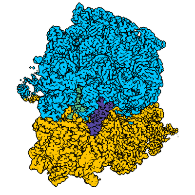 |
Initiation complex consisting of E. coli 70S ribosome with m6AAA mRNA codon in the A-site incubated with a ternary complex containing cognate acylated tRNA(Lys) [9366 multi-frame micrographs composed of 40 frames each in TIFF format] | Koziej L, Glatt S [Pubmed: 37553384] [DOI: 10.1038/s41467-023-40422-7] |
3.3 TB | 2.37 - 2.85 Å |
| 2023-08-18 |  |
Initiation complex consisting of E. coli 70S ribosome with AAA mRNA codon in the A-site [10115 multi-frame micrographs composed of 40 frames each in TIFF format] | Koziej L, Glatt S [Pubmed: 37553384] [DOI: 10.1038/s41467-023-40422-7] |
3.8 TB | 2.04 Å |
| 2023-08-18 |  |
Initiation complex consisting of E. coli 70S ribosome with AAm6A mRNA codon in the A-site [10276 multi-frame micrographs composed of 40 frames each in TIFF format] | Koziej L, Glatt S [Pubmed: 37553384] [DOI: 10.1038/s41467-023-40422-7] |
4.0 TB | 2.04 Å |
| 2023-08-18 |  |
Cryo-EM study on a single, highly heterogeneous cellular fraction with megadalton complexes derived from Chaetomium thermophilum [522 multi-frame micrographs composed of 30 frames each in MRC format] | Semchonok DA, Kyrilis FL, Hamdi F, Kastritis PL [DOI: 10.2139/ssrn.4211492] |
489.4 GB | 3.46 - 3.74 Å |
| 2023-08-18 |  |
Cryo-EM structure of the folate-specific ECF transporter complex in MSP2N2 lipid nanodiscs bound to ATP and ADP (200 kV) [multiple data sets in TIFF format] | Thangaratnarajah C, Rheinberger J, Paulino C, Slotboom DJ [Pubmed: 37491368] [DOI: 10.1038/s41467-023-40266-1] |
580.8 GB | 3.2 Å |
| 2023-08-18 |  |
Cryo-EM structure of the folate-specific ECF transporter complex in MSP2N2 lipid nanodiscs bound to AMP-PNP (200 kV) [3527 multi-frame micrographs composed of 60 frames each in TIFF format] | Thangaratnarajah C, Rheinberger J, Paulino C, Slotboom DJ [Pubmed: 37491368] [DOI: 10.1038/s41467-023-40266-1] |
664.6 GB | 3.6 Å |
| 2023-08-18 | 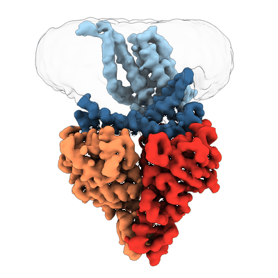 |
Cryo-EM structure of the wild-type solitary ECF module in MSP2N2 lipid nanodiscs in the ATPase open and nucleotide-free conformation (200 kV) [multiple data sets in TIFF format] | Thangaratnarajah C, Rheinberger J, Paulino C, Slotboom DJ [Pubmed: 37491368] [DOI: 10.1038/s41467-023-40266-1] |
1.9 TB | 3.8 Å |
| 2023-08-18 | 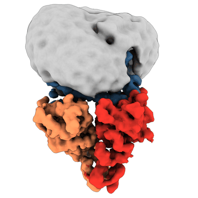 |
Cryo-EM structure of the wild-type solitary ECF module in DDM micelles in the ATPase open and nucleotide-free conformation (200 kV) [1862 multi-frame micrographs composed of 60 frames each in TIFF format] | Thangaratnarajah C, Rheinberger J, Paulino C, Slotboom DJ [Pubmed: 37491368] [DOI: 10.1038/s41467-023-40266-1] |
360.8 GB | 4.3 Å |
| 2023-08-18 |  |
Mouse heavy chain apoferritin in vitreous ice after plunge-freezing [4745 multi-frame micrographs composed of 784 frames each in EER format] | Bongiovanni G, Harder OF, Voss JM, Drabbels M, Lorenz UJ [Pubmed: 37219589] [DOI: 10.1107/S2059798323003431] |
1023.1 GB | 1.61 Å |
| 2023-08-18 |  |
Negative stain EM structure of the NF155 extracellular domain [95 micrographs in MRC format] | McKie SJ, Deane JE [Pubmed: 36996106] [DOI: 10.1073/pnas.2218823120] |
1.5 GB | 19.0 Å |
| 2023-08-18 | 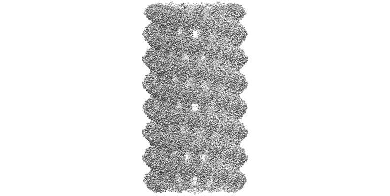 |
KpFtsZ–Mb double helical tube [3096 multi-frame micrographs composed of 60 frames each in TIFF format] | Fujita J, Amesaka H, Yoshizawa T, Hibino K, Kamimura N, Kuroda N, Konishi T, Kato Y, Hara M, Inoue T, Namba K, Tanaka SI, Matsumura H [Pubmed: 37429870] [DOI: 10.1038/s41467-023-39807-5] |
794.4 GB | 2.67 Å |
| 2023-08-18 | 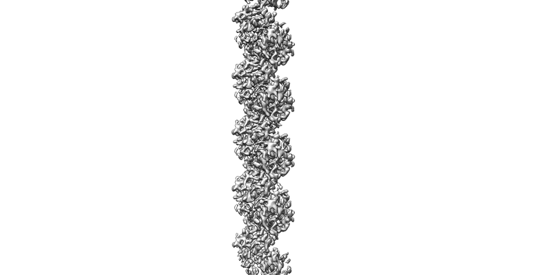 |
KpFtsZ single filament [6079 multi-frame micrographs composed of 60 frames each in TIFF format] | Fujita J, Amesaka H, Yoshizawa T, Hibino K, Kamimura N, Kuroda N, Konishi T, Kato Y, Hara M, Inoue T, Namba K, Tanaka SI, Matsumura H [Pubmed: 37429870] [DOI: 10.1038/s41467-023-39807-5] |
1.7 TB | 3.03 Å |
| 2023-08-18 | 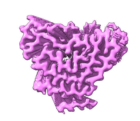 |
Structure of TDP-43 amyloid filaments from type A FTLD-TDP (individual 2) [33336 multi-frame micrographs composed of 40 frames each in TIFF format] | Arseni D, Ryskeldi-Falcon B [Pubmed: 37532939] [DOI: 10.1038/s41586-023-06405-w] |
5.3 TB | 2.39 Å |
| 2023-08-18 | 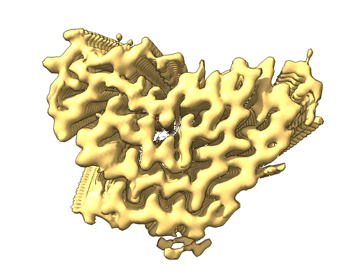 |
Structure of TDP-43 amyloid filaments from type A FTLD-TDP (individual 3) [36507 multi-frame micrographs composed of 40 frames each in TIFF format] | Arseni D, Ryskeldi-Falcon B [Pubmed: 37532939] [DOI: 10.1038/s41586-023-06405-w] |
5.7 TB | 2.39 Å |
| 2023-08-18 | 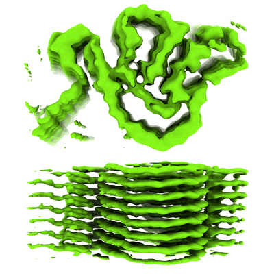 |
Unaligned cryo-EM micrographs of AL55 amyloid fibrils extracted from the kidney of an AL amyloidosis patient [1819 multi-frame micrographs composed of 40 frames each in MRC format] | Chaves-Sanjuan A, Puri S, Schulte T, Ricagno S [Pubmed: 37516426] [DOI: 10.1016/j.jmb.2023.168215] |
1.0 TB | 4.0 Å |
| 2023-08-18 | 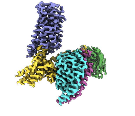 |
Single particle cryoEM of cannabinoid receptor 1/G protein complex bound to an endocannabinoid analogue [8332 multi-frame micrographs composed of 57 frames each in TIFF format] | Kumar KK, Robertson MJ, Skiniotis G, Kobilka BK [Pubmed: 37160876] [DOI: 10.1038/s41467-023-37864-4] |
5.2 TB | 2.8 Å |
| 2023-08-18 | 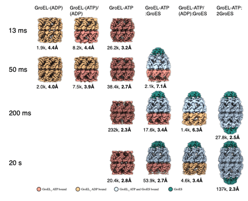 |
Movies of GroEL-ES-nucleotide complex plunged 13, 50, 200 ms and 20 s after mixing with ATP [multiple data sets in TIFF format] | Torino S, Dhurandhar M, Stroobants A, Claessens R, Efremov RG [DOI: 10.1038/s41592-023-01967-z] |
2.9 TB | 2.3 - 7.1 Å |
| 2023-08-18 |  |
Single particle cryo-EM dataset of Mus musculus mitochondrial complex I [2674 multi-frame micrographs composed of 40 frames each in TIFF format] | Grba DN, Chung I, Bridges HR, Agip AA, Hirst J [Pubmed: 37531432] [DOI: 10.1126/sciadv.adi1359] |
1.1 TB | 2.39 Å |
| 2023-08-18 |  |
Single particle cryo-EM dataset of Mus musculus mitochondrial complex I bound with inhibitor Piericidin A [1200 multi-frame micrographs composed of 25 frames each in MRC format] | Grba DN, Chung I, Bridges HR, Agip AA, Hirst J [Pubmed: 37531432] [DOI: 10.1126/sciadv.adi1359] |
1.6 TB | 2.84 Å |
| 2023-08-18 | 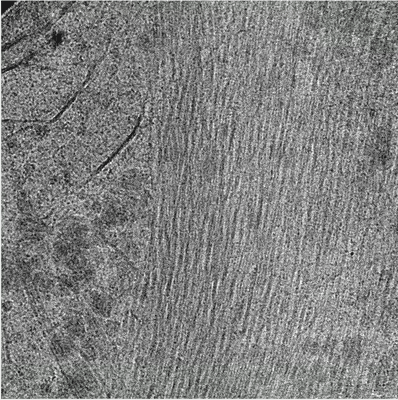 |
Cryo electron tomography of human choriocarcinoma cells [9 multi-frame micrographs composed of 10 frames each in MRC format] | Tun WM, Darrow MC, Basham M [DOI: 10.1017/S2633903X23000107] |
115.3 GB | — |
| 2023-08-18 | 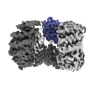 |
AP2 in the presence of heparin [3046 micrographs in MRC format] | Partlow EA, Cannon KS, Hollopeter G, Baker RW [Pubmed: 35347313] [DOI: 10.1038/s41594-022-00749-z] |
267.5 GB | 3.5 - 3.9 Å |
