EMPIAR-11348
Structure of RecT protein from Listeria innoccua phage A118 in complex with 83-mer annealed duplex [2038 multi-frame micrographs composed of 36 frames each in MRC format]
Publication:

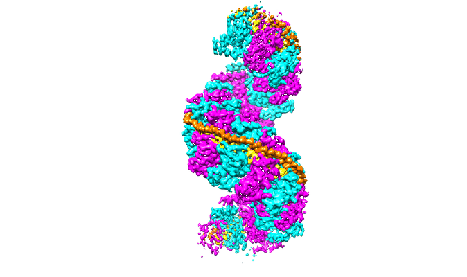
Structure of a RecT/Redβ family recombinase in complex with a duplex intermediate of DNA annealing

Caldwell BJ, Norris AS, Karbowski CF, Wiegand AM, Wysocki VH, Bell CE

Nature communications 13 (2022)

PMID: 36543802

Related EMDB entry:
Deposited:
2022-03-15
Released:
2023-01-30
Last modified:
2023-01-30
Imageset size:
1.73 TB
Imageset DOI:
Experimental metadata:
Download xml json
Contains:
  • micrographs - multiframe
1. Unaligned multi-frame micrographs of LiRecT in complex with 83-mer annealed duplex
Category:
micrographs - multiframe
Image format:
MRC
No. of images or tilt series:
2038
Image size:
(5760, 4092)
Pixel type:
UNSIGNED BYTE
Pixel spacing:
(0.899, 0.899)
Details:
Data collected on 9/17/2020-9/18-2020
Krios, K3, counted mode, EFTEM, Cs corrector
User/Group:Brian Caldwell/Chuck Bell

Image information:
Total # of images = 2038
Total data size = 1.70 TB
Rate of collection = 71/hour
Mode of collection = Accurate hole centering
Gain normalized = YES
File format = mrc
Super-resolution = NO
Spherical aberration = 0.01 mm
Images per hole = 1
Exposure time = 2.7 s
Fractions = 36 [usually 1-2 electrons per fractions]

Microscope parameters:
Pixel size = 0.899 A
Magnification = 81,000
Stage tilt = 0 deg
Dose rate = 19.644 e/px/s
Dose rate = 24.28 e/A^2/s
Total dose = 65 e/A^2 [desired dose rate]
Defocus range = -3.5, -3, -2.5, -2, -1.5, -1
C2 aperture = 50 um
Objective aperture= 100 um
Illuminated area = 1.5 um
Energy Filter = YES
Slit width = 20 eV

Grid type = Quantifoil (gold coated) R1.2/1.3 300 Mesh hole size, mesh, etc.
Position 1 of the large dewar Bell Conical: Position 1 of box J2 (Gray)
Prepared on 8/20/2020 by Brian Caldwell

Additional notes:
Copied gain reference files over into the data folder.
Saved EPU settings file.
Files:
Loading...